


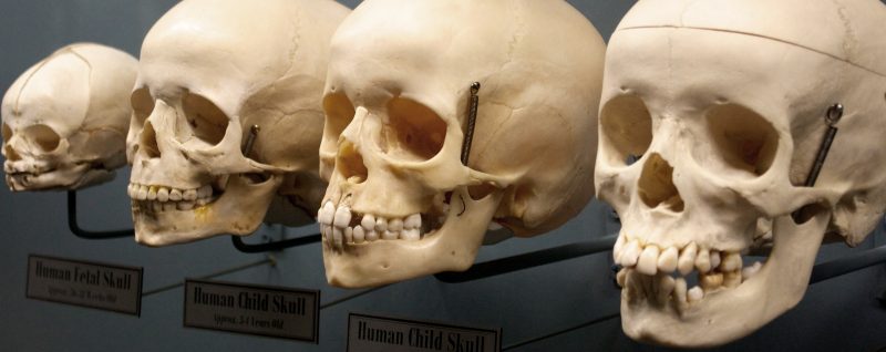
Human beings all develop at generally the same pace but it is not possible to give the exact age of any skeleton – anthropologists can only estimate age at the time death. Size is the major indicator, but once humans reach the age of twelve, skeletal size can be misleading. Looking at the squiggly lines on the skull, called sutures, we try to determine if the suture is completely open, minimally closed, significantly closed or completely closed or fused. Using this information and a scientifically accepted scale we can estimate the age of the owner by how much the sutures have closed since birth. Generally by age 23 the skull sutures have fused closed completely, and by 40 years of age the presence of sutures is barely noticeable.
The size and the shape of the skull will also give us some clear indicators of the origin of the skeleton. Skulls of European ancestry tend to have straight facial profiles and narrower facial surface with projecting, sharply-angled nasal bones. Those from Africa tend to have greater facial projection in the area of the mouth, wider distance between the eyes, a wider nasal cavity, and larger teeth with wrinkling on the molars. Those from Asia have wider facial surface, short, broad cranial vaults with large prominent cheek bones and rounded eye sockets, and large teeth with incisors shaped like shovels.
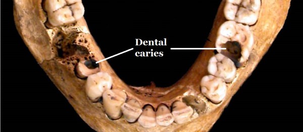
Another helpful hint is in the teeth. We all know that you lose your milk, or baby, teeth at a young age and grow your adult set of dentition afterwards. Identifying the teeth and their size can give fairly accurate pointers to the age of the skeleton. However, once all the adult teeth have erupted, they tend not to be as useful for determining age. But examination of calculus (hardened plaque) on the teeth can show what types of food were eaten by the deceased. Scientists can determine organic as well as non-organic material – whether it was plant or animal, and whether or not it showed signs of cooking – giving us some idea of the diet enjoyed by the owner. Dental caries (or cavities) have become more prevalent over the past few hundred years as our diets slowly changed from whole foods to more processed foods containing greater quantities of sugar. (All Things AAFS)
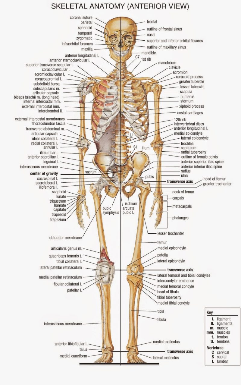
Moving down the body we arrive at the trunk. When a child is born its skeleton is only partly formed and most of the bones are in parts. The epiphyses or ends of the bones and the diaphyses or shafts of the bones develop separately in the womb; when the child is born the bone ends are mostly cartilage with small nubbins of bone starting to form. As we grow, the bone develops and gradually replaces the cartilage, and the shafts get longer. Eventually we end up in our early twenties with a skeleton that will not grow much larger, but with fewer bones than we were born with! So if we want to use the bones to help age the owner we need to look at the development of the bones to give us some starting point. If they have developed completely we then need to look at the degeneration of the bones to see if that helps to determine the age of the skeleton. The joint where the fourth rib meets the sternum is of particular interest, as there are accurate tables that use the amount of cartilage converted to bone at this joint to determine age to a fairly accurate degree. The clavicle is the last bone to finish growing, at around twenty-five years of age. Men tend to have larger muscles than women so the ridges where the muscles attach to the body tend to be larger.
The trunk also contains the pelvis, a vital tool in determining whether the body is of a male or female. Females have a U-shaped sub-pubic arch as opposed to males who show a V-shape. As would be expected, the female’s pelvis is also generally more spacious to allow for childbearing, with a wider sub-pubic angle and sciatic notch. If a woman has given birth there are small pock marks on the inside of the pelvic bone. These marks are caused by the ligaments tearing during childbirth but they only indicate that childbirth has occurred, not how many children were born.
So now we can tell if the owner is a man or a woman and can fairly accurately tell the age and ethnicity. So what else can we learn from the skeleton? We can look for clues to the person’s health. Deficiencies in iron in the owner’s diet can cause swollen marrow spaces in bones. Lack of Vitamin C can cause scurvy (especially prevalent among sailors) and vitamin D can cause rickets – both these diseases cause the bones to soften and bend. Cancerous growths in the soft tissue next to a bone can result in lesions or holes in the bones, while some cancers can cause tumors, resulting in serious deformation of the bones. Infectious diseases also leave tell-tale signs in the bones. Tuberculosis can leave marks on the spine, ribs and pelvis, while syphilis will mark the forehead, nose and shin.
The bones may also tell the story of the person’s occupation. It is well documented that bones change in shape and size in response to what is done to them. If a particular muscle is used often, the bone where the muscle was attached can become thicker, or it may develop ridges in response to the increase in muscle size. This is seen in archers or spear men in ancient times, where repeated throwing or pulling of a bow caused thicker bones at the site of the muscle anchors. Similarly, bones that have atrophied indicate inactivity of the owner. Athletes tend to have deterioration of the hips and knees, and foot soldiers forced to march long distances show a similar pattern of degeneration. Gladiators show excessive trauma to their bones, with breaks and other injuries showing up clearly. Ancient farmers show damage to the shoulders, upper arms and spine as a result of carrying heavy loads for most of their lives, but similar damage can be seen in the skeletons of rowers of ancient galleys. Slaves forced to work in mines show chemical damage caused by working in toxic environments. Of course, all these markers are only indicators to the owner’s life and, on their own, cannot categorically identify the job the owner did.
The work done by anthropologists to understand the life led by the owner of a skeleton is both fascinating and rewarding. No longer is this just a collection of some two hundred and six bones, it defines a living, breathing human being; one who lived, loved, worked, became ill and recovered and eventually died – a person just like me and you. Whether modern or prehistoric, the bones collected can help to make the person real for the researcher.
Lead image source